How to Get Rid of Cherry Angiomas at Home: What Actually Works and Safe Alternatives
No reliable and safe home methods exist for removing cherry angiomas. Trying to remove them by yourself may lead to pain, bleeding, infection, or scarring. Seeing a professional is the safest choice for treating these common skin lesions.
This information is for adults who have noticed cherry angiomas and seek safe ways to manage or remove them. You will learn why home remedies are risky, how to recognize these lesions, and what to expect from professional removal methods. Understanding proper care and avoiding common mistakes will help you make informed choices about your skin health.
Key Takeaways
- Consult a doctor before removing cherry angiomas to confirm the diagnosis and rule out melanoma or other dangerous lesions.
- Cherry angiomas increase with age, affecting about half of adults over 30 and 75% of those over 75 years old.
- Home remedies vary in effectiveness and safety; many lack scientific support and can cause skin irritation or infection.
- At-home devices like the OcuraLife Plasma Pen offer controlled removal with multiple settings but require careful use and follow all instructions.
- Professional removal methods, including laser and electrocautery, provide safer and more precise treatment with faster healing.
- Proper aftercare involves keeping the area clean and avoiding picking to prevent scarring and infection during healing.
Can You Safely Remove Cherry Angiomas at Home?
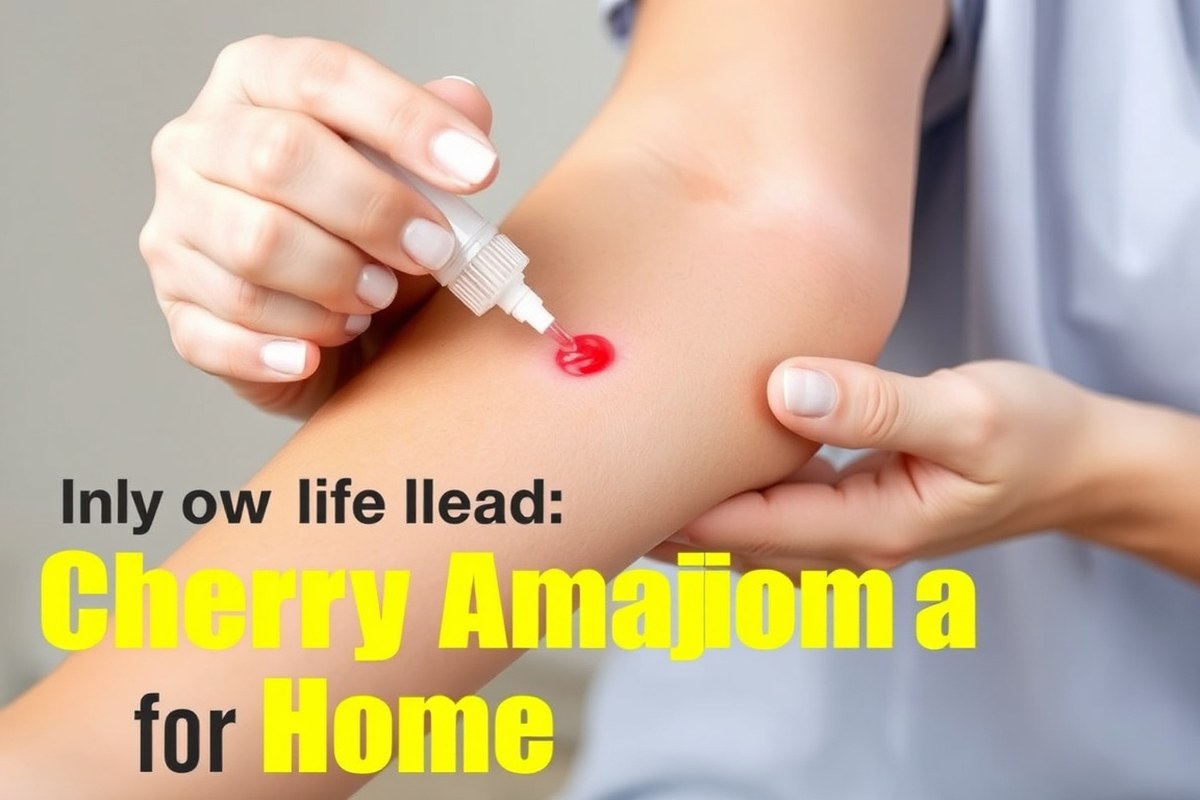
Removing cherry angiomas at home is not safe or recommended. There are no scientifically proven natural or home remedies that effectively remove these small red skin lesions. Attempts to treat them yourself risk pain, bleeding, infection, and permanent scarring. Without proper medical tools and sterile conditions, self-removal often causes more harm than good.
While you might consider using DIY treatments like topical creams, essential oils, or homemade concoctions, none have clinical evidence supporting their effectiveness. These methods may irritate the skin but won’t eliminate the angiomas. Some at-home devices claim to work, such as plasma pens that deliver a focused plasma arc designed for skin lesions, but these should be used only after clear confirmation that the spots are harmless cherry angiomas and not other skin conditions. Improper use increases the risk of skin damage.
Considering action at home, a key decision rule is to avoid any physical trauma to the lesion. Do not try to cut, burn, or freeze cherry angiomas yourself. Professional treatments like electrocautery, laser therapy, cryotherapy, or shave excision are highly effective and safer because they are performed under sterile conditions by trained medical staff who can ensure the lesion is correctly identified and treated. Cost per lesion can add up with professional procedures, but they reduce risks significantly and improve cosmetic outcomes.
For isolated cherry angiomas that appear in visible places without causing symptoms, many dermatologists recommend simply watching them. You should get a professional checkup if the spots change in size, shape, or color, or if they start to bleed on their own. Such exams help exclude serious issues like melanoma.
People with multiple cherry angiomas who want removal might consider visiting a dermatologist to explore options like approved home-use plasma devices, though these should only be used under professional guidance. Such devices may cost several hundred dollars initially but allow treating unlimited spots over time. However, they’re not suitable for all body areas or skin types, so expert advice is critical before starting treatment.
Home removal of cherry angiomas exposes you to risks of infection, scarring, and misdiagnosis. The safest and most effective approach is professional removal, especially when dealing with multiple or changing lesions. Prioritize a dermatologist visit for proper diagnosis and to discuss treatment options tailored to your skin and health history.
Understanding Cherry Angiomas: Causes, Risks, and When to See a Doctor

Cherry angiomas are small, bright red skin growths that many people develop as they age. They are benign and usually don’t cause problems, but understanding their causes and when to seek medical advice helps you manage them safely.
Definition and Appearance of Cherry Angiomas
Cherry angiomas are benign skin lesions made up of clusters of tiny blood vessels. They typically appear as smooth, round or oval spots that range in color from bright red to purple. Usually measuring a few millimeters across, these angiomas can appear anywhere on the body but are most common on the torso, arms, and legs.
Their smooth, shiny surface distinguishes them from other skin issues like moles or skin cancers. Noticing a new red spot that quickly grows or bleeds calls for a medical evaluation.
Genetic and Environmental Causes
The exact cause of cherry angiomas remains unclear, but genetics plays a significant role. They often run in families, suggesting you might be more likely to develop them if your parents have them. Environmental factors like sun exposure have also been linked to their formation.
Other contributors include liver disease and chemical exposure, but these are less common triggers. Avoiding harsh chemicals and excessive sun may help reduce the risk, though there is no certain way to prevent them entirely.
Age and Hormonal Influences
Cherry angiomas are much more common as people get older, especially after age 30. Roughly half of adults over 30 develop at least one, and the number tends to increase with age. Hormonal changes, such as those experienced during pregnancy, can also trigger their appearance.
Women often notice new cherry angiomas after giving birth. The increased levels of certain hormones seem to encourage the growth of these small blood vessel clusters on the skin.
Signs Indicating Medical Evaluation
While cherry angiomas generally don’t present health risks, you should consult a dermatologist if you observe any of the following:
- Sudden eruption of many new angiomas in a short time frame
- Lesions that frequently bleed or are painful
- Rapidly growing spots or changes in size and shape
- Color changes, such as darkening or multiple shades within one lesion
- Spots that ulcerate, crust, or do not heal
- Location in areas that are persistently irritated or injured
Early evaluation helps rule out rare cases of melanoma or other skin cancers that can sometimes resemble cherry angiomas.
Consult your dermatologist promptly if a cherry angioma changes appearance, especially if it bleeds or grows rapidly.
Potential Health Risks and Complications
Cherry angiomas are benign and usually pose no health risks. They rarely bleed unless injured, and they do not turn cancerous. However, repeated trauma to these spots can cause bleeding or infection, which is a reason to protect them from cuts and scrapes.
Some patients may feel self-conscious about the lesions or experience discomfort if angiomas are located where clothing or jewelry frequently rubs against them. In these cases, medical removal options can be considered for comfort or cosmetic reasons.
Common Home Remedies for Cherry Angiomas: Effectiveness and Safety Evaluation
Home remedies for cherry angiomas include various natural oils, extracts, and vinegars, but little scientific evidence supports their effectiveness. These methods generally require consistent application over weeks or months and carry risks such as skin irritation. Knowing the safety concerns and realistic outcomes can help you decide whether to try these treatments.
Topical Oils and Natural Extracts
Castor oil and chamomile oil are commonly applied to cherry angiomas with the hope of shrinking or fading them. These oils have anti-inflammatory properties, but strong evidence that they can remove the lesions is lacking. Applying these oils typically involves gently rubbing the oil on the spot once or twice daily for several weeks using a cotton swab or pad. This method may work better for smaller angiomas and those in sensitive areas, as oils tend to be mild on the skin.
Similarly, iodine ointments are sometimes used for their antiseptic qualities, with some hoping they reduce lesion size. Apply iodine cautiously and only on the angioma, avoiding the surrounding skin to prevent irritation. The total cost for these oils and extracts is typically low, ranging from a few dollars to under $20 per bottle, and they are widely available at pharmacies.
Use of Apple Cider Vinegar
Apple cider vinegar (ACV) is a popular natural remedy for cherry angiomas because it contains acetic acid, which may cause minor skin peeling. Scientific studies do not confirm it can safely or effectively remove cherry angiomas. When using ACV, dilute it with water (one part vinegar to two parts water) to reduce the risk of skin burns. Apply it with a cotton ball for 20 to 30 minutes daily. This treatment needs to continue for several weeks before visible changes might occur, though improvement is not guaranteed.
ACV’s acidic nature can irritate or scar the skin around the angioma, especially if undiluted or left on too long. Costs are low, typically around $5 to $10 per bottle, but you should use caution to avoid skin damage.
Effectiveness of Garlic Applications
Garlic is sometimes recommended due to its natural antibacterial and antioxidant compounds. Some people apply crushed garlic cloves directly to the angioma, covering it with a bandage for 20 to 30 minutes daily. Like other natural remedies, there is no clinical evidence that garlic removes cherry angiomas. The skin can react negatively, showing redness, blistering, or burns, particularly if garlic is applied too long or left on without rinsing properly.
This approach is inexpensive since garlic is common at home but carries a significant risk of skin irritation. Garlic applications can be especially risky on sensitive or thin skin and should be tested on a small area before more extensive use.
Risks and Side Effects of Remedies
Using natural remedies on cherry angiomas can cause skin irritation, redness, burns, scarring, and discoloration. Acidic products like apple cider vinegar or iodine can be harsh and create lasting marks if misused. Applying wart removers or trying to pop angiomas at home increases the risk of infection or bleeding because breaking the skin barrier allows bacteria to enter.
Consult a healthcare provider before starting any home treatment to confirm the diagnosis and rule out other skin conditions. Avoid exposing treated areas to sunlight, which lowers the chance of pigmentation changes. Stop the remedy immediately and seek medical advice if irritation or worsening symptoms develop.
Setting Realistic Expectations for Results
Natural remedies usually work slowly, if at all, requiring daily treatment through several weeks or even months. Most people do not experience significant shrinkage or removal of cherry angiomas from these methods alone. Prepare for minimal visible change, and understand some lesions may remain despite ongoing efforts.
Success varies depending on lesion size, location, and your skin type. Smaller angiomas may respond better to gentle topical oils, while larger or multiple lesions often need professional treatment. Consider home remedies mainly as supportive skin care rather than reliable removal methods.
Comparison of At-Home Devices Versus Professional Removal Methods
When choosing how to remove cherry angiomas at home, the OcuraLife Plasma Pen stands out as the device specifically designed for this type of vascular lesion. It offers single-use tips, 9 adjustable power settings, and detailed instructions, making it the closest match to professional treatments in a home setting. Other common home devices like freezers and generic removal pens are less targeted, which affects effectiveness and safety.
Professional methods such as laser removal, electrocautery, cryotherapy, and shave excision differ significantly in how they target and eliminate cherry angiomas. Laser surgery directs focused light energy to destroy blood vessels underneath the lesion, resulting in precise treatment. Electrocautery uses heat to burn the lesion, which can be effective but may increase the risk of tissue damage. Cryotherapy freezes the lesion, offering a quick option but requiring careful healing management. Shave excision mechanically cuts out raised lesions but involves more procedural effort and downtime.
| Method | How It Works | Main Trade-Off |
|---|---|---|
| Plasma Pen (At-Home) | Produces plasma energy to treat the vascular lesion under the skin | Best home option but limited power and depth compared to professional tools |
| Laser (Professional) | Targets blood vessels with focused light energy to destroy lesion | High precision and good cosmetic results but may not be available at all clinics |
| Electrocautery (Professional) | Uses localized heat to burn and remove the lesion | Effective but increased risk of scarring and burns |
| Cryotherapy (Professional) | Freezes the lesion to cause tissue destruction | Quick treatment but requires careful post-care to avoid irritation |
| Shave Excision (Professional) | Removes raised lesion mechanically by shaving it off | More invasive with longer recovery and possible scarring |
Cost-wise, the plasma pen kit typically ranges from $80 to $150 with one-time use tips, suitable for treating a few angiomas. Professional treatments, depending on the method and clinic, often begin around $150 per lesion, not including follow-up visits. Pain levels with the plasma pen can vary but are generally mild to moderate, often requiring topical numbing. Professional methods usually come with better pain management options but also involve more healing time and occasional side effects like redness or scabbing.
Good results from at-home plasma pens show a clear reduction or disappearance of the cherry angioma after several sessions with proper technique. However, home use carries risks such as ineffective treatment if the energy does not reach the blood vessels below the skin, or skin irritation when power settings are too high. Professional removal methods, especially lasers, tend to provide more consistent outcomes in fewer sessions but at greater cost and clinical supervision.
Professional Cherry Angioma Removal Options and What to Expect
Professional removal of cherry angiomas involves several effective methods including laser treatment, cryotherapy, electrocautery, and shave excision. These options vary in how they work, the level of discomfort involved, and the healing time required. Understanding what to expect can help you choose the best treatment if you opt for professional care over home remedies.
Laser Treatment Details
Laser treatment targets the blood vessels inside the cherry angioma using specific wavelengths, such as pulsed dye or IPL lasers. The heat from the laser causes the tiny blood vessels to collapse and eventually disappear. This procedure is usually quick, often lasting just a few minutes depending on the number and size of lesions. Most patients feel a snapping or stinging sensation during treatment, which is brief and tolerable without the need for anesthesia.
The treated area may appear red or slightly swollen immediately afterward, with mild scabbing that typically clears up within 1 to 2 weeks. Laser removal offers precise results and minimal risk of scarring, making it a good choice for visible areas such as the face or neck. Cost varies widely but can range from $150 to $500 per session depending on location and provider.
Cryotherapy Explained
Cryotherapy uses extreme cold, often liquid nitrogen or a specialized device like the CryoPen, to freeze the cherry angioma. The freezing destroys the abnormal blood vessels beneath the skin surface. This method is straightforward and usually completed in a few seconds to a couple of minutes per lesion. The treatment may cause a brief burning or stinging feeling, but it is generally well tolerated.
After cryotherapy, the treated spot typically forms a blister or scab which falls off in about 7 to 14 days. The skin underneath gradually heals and returns to normal color. Cryotherapy is effective for small to medium-sized cherry angiomas and can be slightly less expensive than laser, often around $75 to $150 per session. Some mild redness or pigment changes can occur but usually resolve over time.
Electrocautery and Shave Excision
Electrocautery involves using a small electric current to burn and seal the blood vessels inside the cherry angioma. This technique generally requires a local anesthetic for comfort because some pain or discomfort can occur during the procedure. It’s a fast treatment and suitable for small angiomas removed in a private clinic setting.
Shave excision physically removes the cherry angioma by shaving off the lesion with a sharp scalpel blade. This method also requires local anesthesia to numb the area, reducing pain during removal. After the procedure, you’ll have a shallow wound that usually starts healing within a few days. Healing can take 1 to 3 weeks depending on size and individual skin response, and dressings may be needed to protect the area initially.
Both electrocautery and shave excision carry some risk of minor bleeding and scarring, especially if the angioma is larger or located on delicate skin. Follow your provider’s instructions on wound care to promote healing and minimize complications. Cost for these procedures can range from $200 to $400 but varies depending on clinic and geographic region.
Always discuss pain management options and realistic healing timelines with your dermatologist before treatment to avoid surprises and ensure proper care.
How to Differentiate Cherry Angiomas from Melanoma and Other Dangerous Lesions
Cherry angiomas are small red or purple growths made up of tiny blood vessels. They most often appear on your torso, arms, or legs and are harmless. However, because they can look similar to serious skin problems like melanoma, it’s important to know the differences and spot any warning signs early.
Melanoma is a type of skin cancer that may appear as a dark spot or mole but can also have red or pink areas, making it tricky to spot. Unlike cherry angiomas, melanoma usually doesn’t blanch, or temporarily fade, when pressed. Cherry angiomas typically do blanch because they are filled with blood vessels that compress under pressure. A simple test is to press on the spot firmly with a clean finger or the edge of a glass. When the redness disappears and returns upon release, it’s likely a cherry angioma. When it stays red or changes in appearance, that’s a reason to seek medical advice. Here is a straightforward checklist to help you perform a self-check for concerning features:
Check size and shape: Cherry angiomas are usually small, round, and uniform, often pinhead-sized up to about a quarter-inch (6 millimeters). Melanomas may have irregular shapes or uneven edges.
Look at color: Cherry angiomas are red to purple and uniform. Melanoma can show a mix of colors including black, brown, tan, pink, or red.
Watch for changes: Track if the spot grows quickly, changes shape, darkens, or bleeds without injury. Such changes require urgent evaluation by a dermatologist.
Notice surface texture: Cherry angiomas are smooth or slightly raised and soft. Melanomas might become scaly, crusted, or ulcerated. Assess symptoms: When the spot itches, hurts, or becomes tender, this is a warning sign. Use this scripted self-check each month:
“I will locate any red or brown spots on my torso, arms, and legs. I will press each spot and note if the redness fades temporarily. Spots larger than a quarter inch, or with irregular borders, multiple colors, or any change from last month, require contacting my dermatologist immediately.”
Medical evaluation is urgent when you notice a spot that doesn’t blanch, bleeds spontaneously, changes size or color quickly, or has irregular borders. Melanomas can develop rapidly and may require biopsy and treatment. Cherry angiomas, by contrast, are benign and pose no health risk unless irritated or bleeding.
Remember, professional skin exams are the safest way to confirm the nature of any suspicious lesion. Dermatologists commonly use tools like dermoscopy to distinguish cherry angiomas from cancer. Having many cherry angiomas or other unusual spots calls for a consultation. The American Osteopathic College of Dermatology also notes that numerous cherry angiomas, especially sudden appearance, can sometimes signal liver problems, so don’t ignore changes in your skin’s pattern.
Proper Care and Healing After Cherry Angioma Removal
After removing a cherry angioma at home, proper care is essential to promote healing and reduce the risk of infection or scarring. The typical healing timeline for a small angioma removed by a minor method such as clipping or cauterization is about one to two weeks. Initially, the treated area might look red and mildly swollen, and a scab will form over the site. Avoid picking or scratching the scab, as this can lead to infection or a permanent scar.
Clean the site gently with mild soap and lukewarm water once or twice a day. After washing, pat the spot dry with a clean towel and apply a thin layer of antibiotic ointment like neomycin or bacitracin. Cover it with a sterile adhesive bandage to protect against dirt and bacteria, especially during the first 48 to 72 hours after removal. Keep the bandage clean and dry, changing it daily or whenever it becomes wet or dirty. When the area is exposed to sunlight, cover it with clothing or use sunscreen after the scab falls off to minimize discoloration or scarring.
Monitoring for complications includes watching for persistent redness beyond a few days, increasing pain, warmth, swelling, or pus, any of which could indicate infection. Severe bleeding that does not stop after gentle pressure and unusual changes like a rapidly growing or bleeding spot also require prompt medical attention. You might notice mild tenderness or itching during healing, that is normal and usually improves over time.
The severity of aftercare depends on how invasive the removal was. Using a sharper tool to excise the angioma can create a slightly larger wound that may take up to three weeks to heal fully. Apply the same cleaning and antibiotic routine, but avoid heavy exercise or activities that stress the treated skin during this longer period. Touching or irritating the site with clothing should be avoided as much as possible.
Proper post-removal care costs little beyond basic supplies like soap, antibiotic ointment, sterile bandages, and sunscreen, often totaling a few dollars. Good results show up as gradual fading of redness and full regrowth of normal skin texture without lumps or discoloration. When scarring occurs, it may take several months to improve. Avoid home remedies that burn or freeze the skin unless designed specifically for cherry angiomas, since over-the-counter freezing kits target warts and skin tags and may injure surrounding tissues.
In all cases, following these care tips helps your skin heal evenly and lowers the chance of complications. Should healing not progress as expected or if you have concerns about the appearance or symptoms, seeking advice from a healthcare professional ensures proper evaluation and treatment options.
Avoiding Common Mistakes When Removing Cherry Angiomas at Home
Trying to remove cherry angiomas yourself often leads to complications such as bleeding, infection, and scarring. Improper techniques or tools can damage the skin and blood vessels, causing not only pain but also longer healing times and unsightly marks. It’s important to recognize when a complication is serious enough to need professional care.
Common mistakes include aggressive scraping or squeezing, which can burst blood vessels and cause heavy bleeding that does not stop easily. Using unclean tools raises the risk of infection, which may cause redness, swelling, pus, or fever. Attempting removal when the spot’s nature is uncertain also risks overlooking a more serious lesion.
- Avoid using sharp objects like needles or razors to cut or puncture the angioma.
- Do not apply harsh chemicals or overheated devices that can burn or irritate the skin.
- Never try to “pop” a cherry angioma as you would a pimple.
- Ensure any tool or product used is sterile and specifically intended for skin treatment.
- Stop immediately if you experience heavy bleeding, spreading redness, or severe pain.
- Do not attempt removal on angiomas larger than 5 millimeters without consulting a doctor.
Watch for signs of complications like bleeding that lasts more than 10 minutes, growing redness or warmth beyond the treatment site, or unusual discharge. These symptoms indicate infection or poor healing and require prompt medical review. If you feel uncertain about the diagnosis or the lesion changes quickly in size or color, seeing a healthcare provider is safer.
Never attempt to remove a cherry angioma if you are not absolutely sure it is benign; a dermatologist can confirm this and recommend safe options.
Stopping home removal attempts at the first sign of trouble is critical. Uncontrolled bleeding or clear infection symptoms need medical attention. Healing without professional input increases the chance of permanent scarring, and only a trained professional can weigh removal benefits against potential risks. Safe removal techniques and proper aftercare can minimize scarring but vary by individual skin type and angioma size.
Step-by-Step Action Plan for Managing Cherry Angiomas at Home
Follow these steps to safely assess, treat, and care for your cherry angiomas while knowing when professional help is necessary.
- Before You Start: Examine the spots carefully and compare them to descriptions of cherry angiomas. Lesions that look irregular, change rapidly, or bleed frequently require a dermatologist visit before attempting any removal.
- Day 1: Choose a safe home remedy or device if you plan removal at home, such as a topical treatment approved for vascular lesions or an over-the-counter freezing kit targeted for skin tags. Make sure to follow the package directions exactly to avoid skin damage.
- Week 1: Apply the chosen home remedy once or twice as directed. Monitor the lesion for changes such as shrinking, color fading, or scabbing, which suggest the treatment is working. Stop use immediately if you experience unusual pain, swelling, or infection signs.
- Week 2: Assess results. When the cherry angioma has not improved or worsens, stop home treatment and book an appointment with a dermatologist to explore professional removal options.
- After Removal (Home or Professional): Keep the treated area clean and dry. Avoid picking or scratching. Use gentle skincare products and apply a healing ointment if recommended. Watch for healing signs like scab formation followed by normal skin reappearance.
- 2 to 4 Weeks Post-Treatment: Check the site for complete healing. Persistent redness, scabbing, or growth should prompt a doctor’s review. Maintain regular skin exams to catch new lesions early.
- Ongoing: Protect your skin from excessive sun exposure to reduce irritation or new lesion formation. Continue observing your skin and consult a dermatologist for any new, unusual, or changing spots regardless of previous treatments.
Frequently Asked Questions
Can You Get Rid of Cherry Angiomas Naturally?
No, there are no proven natural or home remedies to effectively remove cherry angiomas. Professional treatments are the safest option if removal is desired.
Why Do I Have so Many Cherry Angiomas All of a Sudden?
Sudden appearance of multiple cherry angiomas is common as you age, often starting in your 30s or 40s. Hormonal changes and genetics can also play a role.
Do Cherry Angiomas Go Away When Pressed?
Cherry angiomas do not disappear or flatten when pressed because they are clusters of blood vessels under the skin. Pressing them may cause temporary blanching but won’t remove them.
Is There a Cream That Can Remove Cherry Angiomas?
There is no topical cream scientifically proven to remove cherry angiomas. Seeing a dermatologist is necessary for safe removal.
Can Stress Cause Cherry Angiomas to Increase?
Stress does not cause cherry angiomas; they are mostly linked to aging and genetics.
Are Cherry Angiomas Dangerous or Precancerous?
Cherry angiomas are benign and not precancerous; they do not turn into skin cancer.
Key Points to Remember
Safely removing cherry angiomas at home is challenging and often not recommended due to risks of infection, scarring, or mistaking dangerous lesions for benign ones. You can examine your spots closely today using a magnifying glass and check for irregular features like uneven borders or color changes, then consult a dermatologist if anything looks unusual. Scheduling a professional skin evaluation remains the safest step to ensure proper diagnosis and treatment.